
Some post-translational modifications of actin are present in all isoforms (structural variants) of the protein, whereas others are more specific.
The protein’s amino-terminal region can be modified by acetylation (addition of an acetyl group) and arginylation (addition of an arginine amino-acid residue)3. Recent studies identified the enzyme responsible for amino-terminal acetylation of actin and demonstrated that this modification affects the elongation and depolymerization of actin filaments4,5.
Most actin isoforms are also methylated at a particular histidine amino-acid residue known as His73, which is close to the site to which one of two nucleotides, ATP or ADP, binds. Hydrolysis of ATP to ADP plus one free phosphate molecule is essential for the turnover of actin filaments, and hence for their ability to produce force in cells. Although methylation of His73 was identified more than five decades ago6, the enzyme responsible and the biological functions of this modification have remained unknown.
The study by Wilkinson et al. and a related study published in eLife7 report that the SETD3 protein is the enzyme that methylates actin at His73 (Fig. 1). This is the first time an actin methyltransferase (an enzyme that catalyses methylation) has been identified, and also the first time a histidine methyltransferase has been identified in animals. Earlier work suggested that SETD3 methylates lysine amino-acid residues in histone H38, a protein associated with DNA, but Wilkinson et al. convincingly demonstrate that SETD3 is not a methyltransferase for histones. The authors provide extensive biochemical and cell-biological evidence showing that, at least in mammals, SETD3 is the only enzyme that catalyses the His73 methylation of actin, and that actin is the only substrate of SETD3. They also show that SETD3 and His73 methylation of actin are present in a wide range of organisms, including plants and animals, but that SETD3 is not present in budding yeast, which also lacks His73-methylated actin.
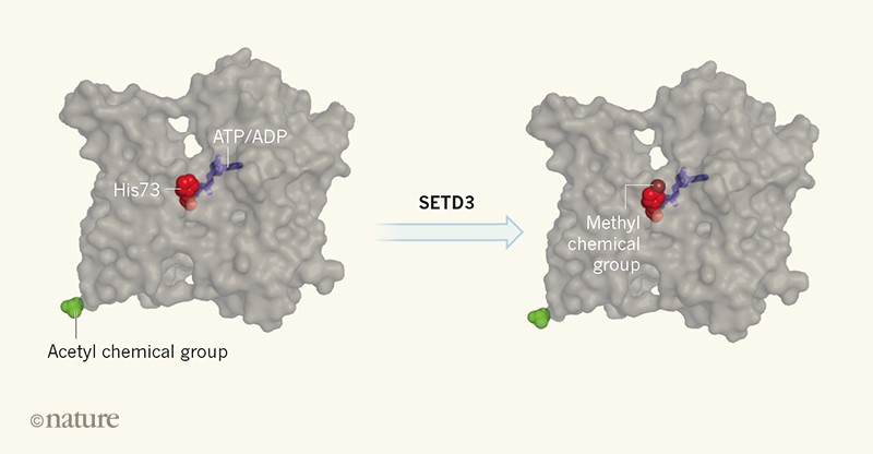
Figure 1 | Methylation of actin by the SETD3 protein.Wilkinson et al.2 show that SETD3 catalyses the addition of a methyl chemical group (methylation) to a histidine amino-acid residue (His73) of the protein actin, and that this modification fine-tunes the protein’s function. His73 is close to a site to which either an ATP or an ADP nucleotide binds. The switch between ATP and ADP is essential to allow actin filaments to produce force in cells. Other evolutionarily conserved post-translational modifications of actin, including the addition of an acetyl chemical group (acetylation) to its amino-terminal region, are distant from the nucleotide-binding pocket of actin.
Why does SETD3 methylate actin, but not other proteins? To answer this question, Wilkinson et al. determined the atomic structure of SETD3 in complex with a short chain of amino acids (a peptide) that has the same amino-acid sequence as the region of actin around His73. They found that this peptide occupies an extended groove in the domain of SETD3 that is responsible for the enzyme’s methyltransferase activity. The interface between SETD3 and the actin peptide has many specific interactions, which explain why SETD3 binds to and methylates only actin.
To examine the biological functions of this post-translational modification, Wilkinson et al. generated ‘knockout’ mice and cell lines in which the gene encoding SETD3 was inactive. They observed that actin is no longer methylated in these models. Surprisingly, the mice lacking SETD3 seemed to be healthy, which demonstrates that methylation of actin at His73 is not essential in mammals. However, female mice lacking SETD3 took longer to give birth than did mice in which this protein was present. The delay resulted from defective contraction of certain muscles of the uterus during labour. Moreover, the migration of SETD3-knockout cells in culture was slower than that of wild-type cells. Finally, non-methylated actin purified from the SETD3-knockout cells polymerized slightly more slowly than did methylated actin, and had a faster rate of exchange of nucleotides on single actin molecules than did actin purified from wild-type cells.
These experiments provide evidence that, despite being evolutionarily conserved across a broad group of organisms, methylation at His73 is not essential for the normal functioning of actin. Instead, this modification seems to fine-tune the protein’s biochemical properties and cellular roles.
Future studies should investigate the SETD3-knockout mice in more detail for possible additional differences from wild-type mice, and should examine the effects of SET
D3 deletion in other model organisms. Also, the effects of His73 methylation on actin biochemistry should be studied more precisely. Previously, analysing these effects was possible only by mutating the His73 residue in actin or by producing human actin in yeast, in which this protein cannot be methylated9,10. The new findings will enable careful side-by-side comparison of wild-type actin and actin that lacks methylation only at His73.
Because His73 is close to the nucleotide-binding site of actin, it will be especially interesting to study how this modification affects the functions of proteins that catalyse nucleotide exchange on actin11and that rely on ATP hydrolysis and subsequent release of free phosphate for their interactions with actin filaments12.
Nature 565, 297-298 (2019)
doi: 10.1038/d41586-018-07882-0


1 Comments
The posttranslational modification of eukaryotic proteins by the addition of isoprenyl lipids at their C-termimi was first observed in the 1970s and 1980s. Since then, Peptide C-Terminal Modification
ReplyDeleteWe will get back to you as soon as possible and thanks for the comment.